• About

Tore L. Hansson

~ Temporomandibular joint dysfunction or craniomandibular disorders from a clinical and practical point of view, focusing on TMJ condylar asymmetry when present.

Tore L. Hansson

Tag Archives: Temporomandibular joint diagnostics

TMJ Condylar Asymmetry

21 Saturday Feb 2015

Posted by torelhansson in Asymmetry Index TMJ, Bruxism, Condylar asymmetry, dentistry, Function & dysfunction of the stomatognathic system, Mandibular position, Orthodontics, Prosthodontics, Retruded Position of the Mandible, Temporomandibular joint (a)symmetry, Temporomandibular Joint Disk Displacement, Uncategorized

≈ 1 Comment

Tags

Arthrogeneous origin of TMJ pain, Asymmetry, Asymmetry Index, Centric relation, condylar asymmetry recognition, Dentistry, Oral rehabilitation, Oral Stability, Orthodontics, Retruded Position of the Mandible, Temporomandibular joint diagnostics, Temporomandibular joint disorder, TMJ rehabilitation, Treatment planning

when present,is a must to consider, in any dental rehabilitation. I dare to have this opinion after more than three decades of experience in treating ” asymmetrical ” patients shared with several successful colleagues. Although I repeat my opinion   it seems relevant as we on a regular basis are exposed to excellent crowns, bridges,supra constructions on implants and even completed orthodontics and yet a not satisfied patient.The chosen mandibular position for the rehabilitation in the majority of these patients has not been correct.

Results of recent research performed in different countries indicate that the mechanics of the temporomandibular joint is essential in order to maintain a pain free and functioning stomatognathic system (Quintessence International Symposium, Scottsdale,Arizona February 6-7, 2015). Overloading of the joint seems  not only to jeopardize the intraarticular structures of the joint resulting in anything from internal derangement to osteoarthritis but also to be the trigger for masticatory muscle pain.

At a vertical temporomandibular joint condylar asymmetry the loading of the two joints is in danger as the vertical dimensions of the two condyles are not equal. Therefor the vertical dimensions of the two temporomandibular joint condyles need to be analyzed before any treatment is initiated. It is of utmost importance to determine the highest condyle as at an asymmetry  this condyle has to be the guide for the mandibular movement of  rotation(the Retruded Position of the Mandible)in which the rehabilitation is going to be executed.

Additionally,in patients with functional facial pain it sometimes might be difficult to clinically manipulate the mandible into the correct position for rehabilitation. At such occasions the result of the vertical condylar analysis in the panoramic X-ray easily can be transferred into the Maaxloc device by Dentatus, in which the index for the mandibular position of the planned rehabilitation is made.

Advertisement

TMD 2015, Condylar Asymmetry?

10 Tuesday Feb 2015

Posted by torelhansson in Arthrogeneous origin of pain, Asymmetry Index TMJ, Bruxism, Condylar asymmetry, Function & dysfunction of the stomatognathic system, Functional pain, Mandibular position, Retruded Position of the Mandible, Temporomandibular Joint Clicking and Popping, Temporomandibular Joint Disk Displacement, Temporomandibular Joint Sounds, Treatment planning, Uncategorized

≈ 2 Comments

Tags

Arthrogeneous origin of TMJ pain, Asymmetry, Asymmetry Index, Bruxism, condylar asymmetry recognition, Dentistry, Occlusal device, Retruded Position of the Mandible, Temporomandibular joint diagnostics, Temporomandibular joint disorder, Temporomandibular joint pain, TMJ rehabilitation, Treatment planning

Reflecting back on the interesting information given by eighteen well known international speakers at the Quintessence International Symposium on TMD & FACIAL PAIN and HEADACHE, Bridging the Gap Between Current Research and Clinical Practice in Scottsdale Arizona ( February 6-7,2015, Co-Chaired by Drs Daniel M.Laskin and Charles S.Greene ) I once again would like to stress the importance to recognize a temporomandibular joint condylar asymmetry when present. Several speakers stressed the overloaded temporomandibular joint as the cause of TMD and advised to pay less attention  to the masticatory muscles initially at  the examination of TMD pain. Lack of synovial liquid and mini ruptures or even nano ruptures of the joint tissues are to be looked upon as causes for cartilage degeneration with osteoarthritis / osteoarthrosis as the end result.

It is my opinion that in combination with bruxism a temporomandibular joint condylar asymmetry needs to be acknowledged and addressed. The shorter condyle side joint is in its morphology unstable and easily develops internal derangement of its articulating parts.  The well known phenomenon of disk displacement is thereby a fact and the first step in joint break down with the diagnosis Arthritis microtraumatica art.temp.mand. In this perspective occlusion becomes extremely important . The shorter condyle side joint needs to be supported with the correct vertical dimension of occlusion. What is missing in vertical dimension inside that joint needs to be compensated for at the level of the teeth of the ipsilateral side. In my mind this is the only possibility to create a stable occlusion with the maintenance of improved joint function after the initially reversible approach with an orthotic device.

Finally a statement/question that might need some thoughts: How much longer are we as a profession going to talk about occlusion without considering the vertical dimension of the two temporomandibular joints!?

House of Gnathology?

13 Monday Oct 2014

Posted by torelhansson in Arthrogeneous origin of pain, Asymmetry Index TMJ, Condylar asymmetry, dentistry, Function & dysfunction of the stomatognathic system, Functional pain, Mandibular position, Myogeneous origin of pain, Occlusal adjustment, Occlusion, Orthopantomogram OPG, Prosthodontics, Temporomandibular joint (a)symmetry, Temporomandibular Joint Disk Displacement, Tension headache, tooth clenching, tooth grinding, Treatment planning, Uncategorized

≈ Leave a comment

Tags

Arthrogeneous origin of TMJ pain, Asymmetry, Bruxism, Centric relation, condylar asymmetry recognition, Dentistry, Myogeneous origin of TMJ pain, Occlusal adjustment, Oral Stability, Retruded Position of the Mandible, Temporomandibular joint diagnostics, Temporomandibular joint disorder, Temporomandibular joint pain, TMJ rehabilitation, Treatment planning

A week ago I had the pleasure to attend the XXVII International AIG (Associazione Italiana di Gnatologia) Congress in Torino, Italy. Twentyseven speakers from six countries each had about 30 minutes to present their thoughts about the relationship between occlusion and temporomandibular joint disorders. In general it could be concluded that a stable occlusion would improve the function of the stomatognathic system and thereby reduce the risk for developing or aggravating a temporomandibular joint disorder. It was also underlined in several lectures that gnathology is a team work and that occlusion is one tool in the treatment of a temporomandibular joint disorder.
It was interesting to note that the speakers in general agreed in finding occlusion an important factor in the treatment of a temporomandibular joint disorder, i.e. a complete contradiction to the conclusion of several hundreds of articles and studies proclaiming that when RDC/TMD are used in the evaluation of treatment outcome occlusion is not important and should not get priority or most often not even be considered in the treatment of a temporomandibular joint disorder.

RDC/TMD were discussed and by a great part of the participants even questioned in the final round table discussion of the congress. In my opinion and in agreement with many other clinicians RDC/TMD cannot and should not be used in the clinical situation in which a specific diagnosis is to be the sound base for the treatment. Reliable criteria like maximum mouth opening, mandibular movements, the difference between dynamic and static pain and even the difference between the vertical dimensions of the two temporomandibular joint condyles visible and measurable in a panoramic X-ray exist. Therefore my suggestion to the RDC/TMD supporters is: Next time you conduct a clinical study, why not incorporate the above mentioned criteria! I am pretty convinced that the results will be different regarding the eventual link between occlusion and the temporomandibular joint disorder.

Finally I would delete the ? from the title of this blog and agree with the suggestion from Dr.Eugenio Tanteri that a forum “House of Gnathology” might come to an agreement that a correctly designed and applied occlusion is important in providing  oral stability and thereby reduce the prevalence of temporomandibular joint disorders among our patients.

Asymmetry Index ( AI ) for temporomandibular joint Condylar Asymmetry

26 Wednesday Feb 2014

Posted by torelhansson in Arthrogeneous origin of pain, Asymmetry Index TMJ, Condylar asymmetry, dentistry, Function & dysfunction of the stomatognathic system, Functional pain, Mandibular position, Myogeneous origin of pain, Occlusion, Prosthodontics, Retruded Position of the Mandible, Temporomandibular joint (a)symmetry, Tension headache, Treatment planning, Uncategorized

≈ 2 Comments

Tags

Arthrogeneous origin of TMJ pain, Asymmetry, Asymmetry Index, Centric relation, condylar asymmetry recognition, Dentistry, Myogeneous origin of TMJ pain, Occlusal adjustment, Oral rehabilitation, Oral Stability, Retruded Position of the Mandible, Reversible treatment, Temporomandibular joint, Temporomandibular joint diagnostics, Tension headache, TMJ rehabilitation

For more than twenty years I have been working on a regular basis with Dr Ugo Gambardella, Medical Space in Seriate, Italy. In 1993 we founded the T.A.C. Seriate clinic, ( Team Approach to Craniomandibular disorders ) in order to optimize the diagnostics and treatment of patients suffering facial pain due to dysfunction of the stomatognathic system. Dr Gambardella was already then experienced in prosthodontics as well as in implantology.
Already at the start it became obvious to us that a large portion of the mostly referred patients showed a condylar asymmetry and a lack of oral stability. Initially our treatment aimed at a normalization of the stomatognathic system via a stabilization splint in its design compensating for the condylar asymmetry, when present, and providing at least a temporary oral stability. After 6 – 12 months several patients wanted to have permanent oral stability, which then was achieved through mostly prosthodontics , sometimes in combination with preprosthetic orthodontics.
Some years ago and then more than 5 years after completed treatment the patients were invited to have their situation evaluated and to tell how they felt and how they were functioning after treatment. The equivalent clinical examination of their stomatognathic system was executed by an Italian speaking colleague, who did not know what kind of treatment the patients had got or been exposed to. The teeth depicted in the panoramic overview X-rays were covered i.e. made not visible to the examiner while the two temporomandibular joints were shown.
The condylar asymmetry was analyzed and for each patient an Asymmetry Index ( AI ) was calculated according to the following formula: ( R – L : R + L ) x 100 i.e. the right side condylar height minus the left side condylar height divided by the sum of the right and left side condylar heights multiplied by 100. Thus an asymmetry index was expressed in percentage for each patient.
The results of the subsequent analysis showed that after more than five years with permanent oral stability no patient any longer was found with a mainly arthrogeneous origin of pain. Myogeneous origin of pain was also frequently eliminated or markedly reduced. All patients showed an increase of maximum mouth opening.
The patients were grouped and analyzed according to the asymmetry index ( AI ) . It can be concluded from the results that patients with an AI > 3 % in general benefit from reversible treatment. Patients with an AI in the range between 3 % and 10 % may do well with just a temporary oral stability in form of a stabilization splint i.e. the splint can be worn part time, which for most patients means during sleep. As the participating patients with an AI > 10 % definitely benefitted the most and also felt subjectively completely different, mostly totally pain free after the treatment it is our opinion, that when you find a condylar asymmetry > 10 % in a patient with a diagnosed facial pain, you can comfortably suggest permanent occlusal treatment after the initially reversible period in the correct mandibular position guided by the higher temporomandibular joint condyle. The permanent treatment then executed in the same mandibular position will provide oral stability.

TMJ Clicking Sound

10 Monday Feb 2014

Posted by torelhansson in Arthrogeneous origin of pain, Bruxism, Condylar asymmetry, dentistry, Function & dysfunction of the stomatognathic system, Functional pain, Mandibular position, Myogeneous origin of pain, Occlusion, Retruded Position of the Mandible, Reversible treatment, Temporomandibular joint (a)symmetry, Temporomandibular Joint Sounds, Uncategorized

≈ 2 Comments

Tags

Arthrogeneous origin of TMJ pain, Asymmetry, Centric relation, condylar asymmetry recognition, Dentistry, Myogeneous origin of TMJ pain, Retruded Position of the Mandible, Temporomandibular joint, Temporomandibular Joint Clicking, Temporomandibular joint diagnostics, Temporomandibular Joint Disk Displacement, Temporomandibular joint pain, Temporomandibular Joint Sounds

TMJ clicking sound has been reported, discussed, investigated and even visualized in thousands of articles during the past thirty and more years with a peak of interest in 1980-s. Common knowledge is that the sound is either due to deviations in form of the articulating surfaces of the condyle and / or the temporal component ( increase of cartilage in specifically overloaded areas of the articulating surface ) or to a displacement of the disk. Disk displacement often comes and goes and it is therefore not a surprise that it is more common among patients with a mainly myogeneous origin of pain. Since the result of comprehensive epidemiological  studies in the 1970-s it is also common knowledge that  the phenomena occurs in about 40% of the adult population.

Hopefully in not contributing to the too often clinically existing confusion of how to handle the sound I would rather suggest the following: Check the panoramic x-ray i.e. check specifically the temporomandibular joint condylar heights before you take any further steps in your attempt to help your patient!

Most probably you will find a shorter condyle of the joint with the registered clicking sound. Try to guide a mandibular rotation movement around the higher condyle and most likely the clicking sound disappears or at least gets a reduced  magnitude, when it is due to a latent disk displacement. If there is no discomfort or pain to be registered there is no real indication for intervention. However, if discomfort or a mainly myogeneous origin of pain is reported by the patient there is a need for an initially reversible treatment in form of a stabilization splint that compensates for the difference in vertical dimensions between the two condyles. The splint is to be worn part time . The patient is supposed to find a seemingly fast relief.  When the clicking sound is found in combination with a mainly arthrogeneous origin of pain the splint needs to be worn as much as possible for at least 6 months before additional permanent treatment is considered.

In addition , even if the clicking sound is due to a deviation in form ( of the condyle and / or of the temporal component )  the clicking sound may over time get reduced as a smoothening out of the articulating surfaces can be expected at a consequent long term  wearing of the correctly designed splint. Therefore I once again would suggest the use of the Maaxloc instrument in producing the index  which will be used in mounting the models into the articulator before the construction of the stabilization splint. The splint absolutely must compensate for the registered difference in vertical condylar height between the two joints in providing a symmetrical oral stability in order to eliminate the  painful and bothersome  tmj clicking.

Sleep Apnea Oral Devices

05 Wednesday Feb 2014

Posted by torelhansson in Arthrogeneous origin of pain, Condylar asymmetry, dentistry, Function & dysfunction of the stomatognathic system, Functional pain, Mandibular position, Myogeneous origin of pain, Occlusion, Orthopantomogram OPG, Reversible treatment, Temporomandibular joint (a)symmetry, Uncategorized

≈ Leave a comment

Tags

Arthrogeneous origin of TMJ pain, Asymmetry, condylar asymmetry recognition, Dentistry, Myogeneous origin of TMJ pain, Occlusal device, Sleep Apnea, Sleep Apnea Device, Temporomandibular joint, Temporomandibular joint diagnostics, Temporomandibular joint dysfunction, Temporomandibular joint pain, Treatment planning

Nowadays MAD, the mandibular advancement device, seems to be delivered from almost every dental office in an effort to assist the medical profession fighting sleep apnea, a major problem for many of our patients. The device is supposed to direct the mandible forward and inferiorly in order to increase the air flow through the mouth. However, a standardized method in making the device seems lacking and many patients suffer discomfort or pain after wearing the device after various lengths of time. Dentists regularly also register changes of occlusion, most often posterior open bites.

As the device shall position the mandible forwards the most often clinically achieved index accompanying the impressions to the laboratory must be taken in a protruded mandibular position. But how much protrusion seems to be left at random. There are studies concluding that it does not matter whether the protrusion is 75% of maximum protrusion or only 25%. Nevertheless it seems accepted to allow a rather large amount of protrusion as additional protrusion as adjustment of the device  often is recommended in the cases where the originally designed device has not given the expected relief.

At a vertical temporomandibular joint condylar asymmetry an excessive and prolonged protrusion definitely puts the comfort of the patient and a  harm free function of the stomatognathic system at risk. At a vertical condylar asymmetry most likely the clinically taken index does not register the mandible in a symmetrically protruded mandibular position. Most likely the mandible is deviating towards the ” shorter ” condyle side. To then have this position maintained during six to eight hours of sleep with thus an awkward  and asymmetrical muscle function and temporomandibular joint condylar positions may be one reason for discomfort or facial pain at awakening.

One way to avoid the problem is to use the Maaxloc ( following the analysis of the vertical condylar heights in the panoramic x-ray ) in constructing the index which is to be used in the mounting of the models in the articulator. It is then easy to bilaterally just set the amount of individually determined protrusion to be applied in the fabrication of the device. A symmetrical protrusion is thereby achieved resulting in a more comfortable device.

A good friend of mine, Dr Staffan Lindholm, Stockholm, Sweden, is probably one of the most experienced dentists in Scandinavia when it concerns oral devices  in fighting sleep apnea. His expertise and unusual self criticism already several years ago  motivated him to compare the outcome of 59 conventionally and from just clinically taken indices  oral devices with another  59 devices constructed from articulator set protrusion of the models mounted with the Maaxloc registration. Six patients of the first group suffered pain and discomfort from  their devices while only 1 patient of the second group showed a minor dissatisfaction, but tolerated the splint. The 6 not successful devices in the first group of  patients were later adjusted according to a Maaxloc registration resulting in patient satisfaction.

In many discussions with fellow dentists and dental technicians Dr. Lindholm  proclaims the superiority of the Maaxloc instrument in constructing a more comfortable  and successful oral device. He is happy to generally tell about his continuing success with a 80-90% reduction of the risk to develop discomfort or pain in the patients who are forced to wear an oral device fighting sleep apnea by using the Maaxloc registration in mounting the models before its fabrication in the laboratory . In conclusion, please do consider a correct index in protrusion for the construction of any anti sleep apnea device at your choice and thereby enjoy a major increase of clinical success!

Maaxloc, the mandibular axis locator

07 Tuesday Jan 2014

Posted by torelhansson in Abused protrusion, Arthrogeneous origin of pain, Bruxism, Function & dysfunction of the stomatognathic system, Functional pain, Mandibular position, Myogeneous origin of pain, Occlusion, Orthopantomogram OPG, Retruded Position of the Mandible, Temporomandibular joint (a)symmetry, Temporomandibular joint morphology, Tension headache, TMJ radiology, tooth clenching, tooth grinding, Uncategorized

≈ 4 Comments

Tags

Asymmetry, Bruxism, Centric relation, condylar asymmetry recognition, Condyle (anatomy), Dentistry, Occlusion, Radiography, Retruded Position of the Mandible, Temporomandibular joint, Temporomandibular joint diagnostics, Temporomandibular joint disorder, Temporomandibular joint pain

photo-17Several years ago I met with and was introduced to Kenneth Wallenholm, an experienced dental technician in Stockholm, Sweden. I was giving a lecture and he was in the audience. I had talked about the recognition of temporomandibular joint condylar asymmetry, when present, and its importance for a correct registration of the mandibular position to be used as the reference position for optimal dental work clinically as well as in the dental laboratory. He had immediately understood my message and for a long time also realized that most “wax bites”, indices or ” Registrat ” accompanying dental impressions into the laboratories most frequently ended up in the trash can, never being used. The reason was that these ” wax bites ” did not provide any information about the workable intermaxillary relation. The technicians therefore rather related the upper and lower models of the jaws by hand i.e. they just tried to find the best stability between the models,maybe sometimes the intercuspal position, before they started their technical procedures to establish an acceptable occlusion. We were  both of the same opinion that such unfortunate events outside the mouth of the patient are the major reasons  for clinically unwanted adjustments of dental work before its  insertion into the mouth.

It did not take us long time until we met again and started to brain storm how to come up with a device in order to facilitate for the dentist / technician team to ascertain and improve the registration of the correct mandibular position to be used by both the professions. After many hours, many different measurements, several different drawings and a certain number of pieces of ” metal constructions ” the patent was approved and the prototype for the Maaxloc  was completed by Dentatus ( info@dentatus.se : dentatus@dentatus.com )

The Maaxloc determines the clinically correct mandibular position. The Maaxloc is not an articulator even if it is described under articulators in the list of products from Dentatus. The Maaxloc is a device to assist in finding the correct mandibular position of reference at a recognized condylar / mandibular asymmetry. The registration index produced in the Maaxloc can be  used in any articulator of your choice and can comfortably be applied in cases of craniomandibular or temporomandibular disorders,as well as in any case of prosthetic rehabilitation or orthodontic analysis. Determining the correct mandibular position of reference is of utmost importance and will help to avoid unwanted occlusal features. This includes achieving the correct incline of implants into the jaws and improving the occlusal analysis before an orthodontic treatment is initiated.

The Maaxloc is time saving and reduces pain because:

1.Symmetry in occlusal contacts between the jaws decreases the risk of failure in providing bilateral load to the stomatognathic system.

2.Less time is spent on adjustments and on remakes due to fractures in veneers and crown & bridge work, including supra structures on successfully accepted implants.

3.Shorter sessions for repetitive registrations of the mandibular positions.

4.A stable occlusion compensates a morphological asymmetry, improves the general adaptation of tissues and is especially beneficial to the edentulous patient wearing removable prostheses, with less dislocation of the prostheses.

5.Last but not least, once the individually correct mandibular position has been found, the patients immediately acknowledge their natural way of achieving and sensing oral stability

It  is my convinced opinion that the measurements of the condylar heights in a panoramic ( overview ) x-ray and  the difference between the two sides transferred into  the Maaxloc will further your clinical success and confidence.

The Mandibular Position and The Hinge Axis

06 Monday Jan 2014

Posted by torelhansson in Arthrogeneous origin of pain, Function & dysfunction of the stomatognathic system, Functional pain, Mandibular position, Temporomandibular joint (a)symmetry, Temporomandibular joint morphology, TMJ radiology, Uncategorized

≈ Leave a comment

Tags

Asymmetry, Bruxism, Centric relation, condylar asymmetry recognition, Condyle (anatomy), Dentistry, Radiography, Retruded Position of the Mandible, Temporomandibular joint diagnostics, Temporomandibular joint disorder, Temporomandibular joint pain

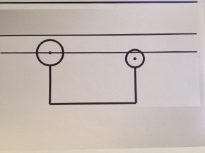
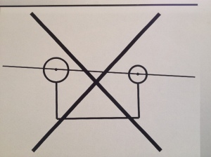
Correct axis of rotation at condylar asymmetry It is impossible for me not to repeat the importance in every clinical situation to recognize a temporomandibular joint condylar asymmetry, when present. In not doing so the treatment cannot be correctly predicted. The treatment will not be as successful as was expected . Unnecessary adjustments of oral devices or, even worse, adjustments of permanent dentistry will disappoint the patient as well as the dentist with also the risk that the treatment will never be fully accepted despite tremendous efforts from both parties involved.

In the clinical situation or in the dental laboratory the conventional axis of mandibular rotation should never penetrate the centers of the two temporomandibular joint condyles at a condylar asymmetry.One condyle, the ” higher one “must determine the axis of rotation. If not, the axis of rotation could not be parallel to the horizontal plane, used as the reference at general rehabilitation.

image-1                                                  Wrong axis of rotation at a condylar asymmetry.

Correct axis of rotation at condylar asymmetryCORRECT AXIS OF ROTATION AT A CONDYLAR ASYMMETRY.

It is my opinion that the definition and the application of the generally used retruded position of the mandible ( RPM ),based upon the assumption that the two temporomandibular joints are equal, therefore needs to be reconsidered and slightly adjusted Clinically I have always worked with the retruded position of the mandible as my reliable position of reference.However for the past 25 years I  have advocated that the retruded position of the mandible ( RPM ) is ( should be ,if not yet ) determined and guided by the normally developed and functioning temporomandibular joint condyle. When the right side is the higher side RPM is to be written RPM (R). When the left side is the higher side RPM is to be written RPM (L) and when the two condyles are of equal height RPM is to be written RPM (=).In addition, when the two condylar heights are measured in the panoramic ( over view ) x-ray and the magnification factor  of the machine is compensated in the calculation the RPM(R) or RPM(L) at an asymmetry can be written i.e. RPM(R)1,5mm or RPM(L) 0,5 into the clinical documents. In that way the almost true difference between the two condylar heights easily can be compensated for in the necessary index to be used in the phase of rehabilitation clinically or at the laboratory via the Maaxloc device (info@dentatus.se)

In general the higher condyle determines what side is to be the guiding side for RPM. However, there is one exception under rare circumstances.At a ramus asymmetry when the total vertical dimensional difference between the two mandibular sides is determined by a major difference in height of the two rami, the vertical dimensional difference determined by the condyles is overruled.Then the biggest total mandibular vertical height may direct even the “shorter condyle” to be the guide in the registration of the retruded position of the mandible and the center of the hinge axis rotation. In this context “disturbing” precontacts or interferences enter a completely new perspective. Unilateral tooth contacts on the “higher condyle” side may indicate the correct vertical dimension of the occlusion. The lack of tooth contacts on the “shorter condyle” side may indicate the occlusal dimension that needs to be increased.Only interferences or precontacts on the “shorter condyle” side will then remain as traditional interferences i.e.disturbing tooth contacts.

Team Approach in tmj Condylar Asymmetry Recognition and rehabilitation( TACAR )

01 Sunday Dec 2013

Posted by torelhansson in Function & dysfunction of the stomatognathic system, Temporomandibular joint (a)symmetry, Temporomandibular joint morphology

≈ 1 Comment

Tags

condylar asymmetry recognition, Temporomandibular joint diagnostics

For many years during and after given lectures and courses I have been asked to guide and to assist in the diagnostics and treatment of facial pain and often facial asymmetry due to a vertical temporomandibular joint condylar asymmetry.Often dentists do not consider the phenomenon, a possible vertical temporomandibular joint condylar asymmetry, when their utmost perfect occlusal rehabilitation fails. It is not that strange as mostly the two temporomandibular joints were not considered in the planning and design of the prosthodontics or orthodontics.There may be several explanations and  reasons.Some of them may be ascribed to facts like:

1. The temporomandibular joints are often pictured in a sagittal plane and one at the time,

2.The two temporomandibular joints are learnt and conceptualized as being identical,

3.An occlusal adjustment is something that is considered almost “normal “, when ” the work ”  returns from the dental laboratory before it’s permanent insertion into the mouth,

4.Occlusal devices need to be adjusted as the patient might be a tooth grinder or a tooth clencher,

5.The two temporomandibular joints may not be or were not visible in the panoramic radiograph (if even taken ) or

6.The dentist was taught that the panoramic radiograph is not reliable and that all dimensions in the pictureare are not trustworthy and so on.

From January 2014 on I will try to provide an exchange of experience and ideas through this blog on at least a weekly basis. The blog will address clinical and practical problems . My intention is to share my experience of more than 30 years in dealing with tmj condylar asymmetry.The readers are most welcome to ask questions and hopefully the blog will stimulate to new discoveries and even to convince the very conservative part of the dental profession to recognize and to compensate for an existing temporomandibulr joint condylar asymmetry when it is confirmed to be the cause for the facial pain in the patient.

Recent Posts

  • House of Gnathology part II
  • Splint (Oral Orthosis) Adjustment, A Necessity?
  • Too many adjustments on the dental work?
  • Prefabricated occlusal devices,mouth guards or splints
  • Splint adjustments

Recent Comments

torelhansson on House of Gnathology part …
Paul C. Riley, DDS on House of Gnathology part …
torelhansson on Asymmetry Index ( AI ) for tem…
R on Asymmetry Index ( AI ) for tem…
marjan on TMJ Condylar Asymmetry

Archives

  • December 2016
  • January 2016
  • December 2015
  • September 2015
  • August 2015
  • July 2015
  • March 2015
  • February 2015
  • December 2014
  • October 2014
  • June 2014
  • May 2014
  • April 2014
  • March 2014
  • February 2014
  • January 2014
  • December 2013

Categories

  • Abused protrusion
  • Arthrogeneous origin of pain
  • Asymmetry Index TMJ
  • Bruxism
  • Condylar asymmetry
  • dentistry
  • Function & dysfunction of the stomatognathic system
  • Functional pain
  • Gnathology
  • Implantology
  • Mandibular position
  • Myogeneous origin of pain
  • Occlusal adjustment
  • Occlusal device
  • Occlusal splints
  • Occlusion
  • Occlusion
  • Orthodontics
  • Orthopantomogram OPG
  • Orthosis
  • Prosthodontics
  • Retruded Position of the Mandible
  • Reversible treatment
  • Temporomandibular joint (a)symmetry
  • Temporomandibular Joint Clicking and Popping
  • Temporomandibular Joint Disk Displacement
  • Temporomandibular joint morphology
  • Temporomandibular Joint Sounds
  • Tension headache
  • TMD
  • TMJ radiology
  • tooth clenching
  • tooth grinding
  • Treatment planning
  • Uncategorized

Meta

  • Register
  • Log in
  • Entries feed
  • Comments feed
  • WordPress.com

Blog at WordPress.com.

Privacy & Cookies: This site uses cookies. By continuing to use this website, you agree to their use.
To find out more, including how to control cookies, see here: Cookie Policy
  • Follow Following
    • Tore L. Hansson
    • Already have a WordPress.com account? Log in now.
    • Tore L. Hansson
    • Customize
    • Follow Following
    • Sign up
    • Log in
    • Report this content
    • View site in Reader
    • Manage subscriptions
    • Collapse this bar
 

Loading Comments...